Structure Parts and Functions of Bacteria Cell
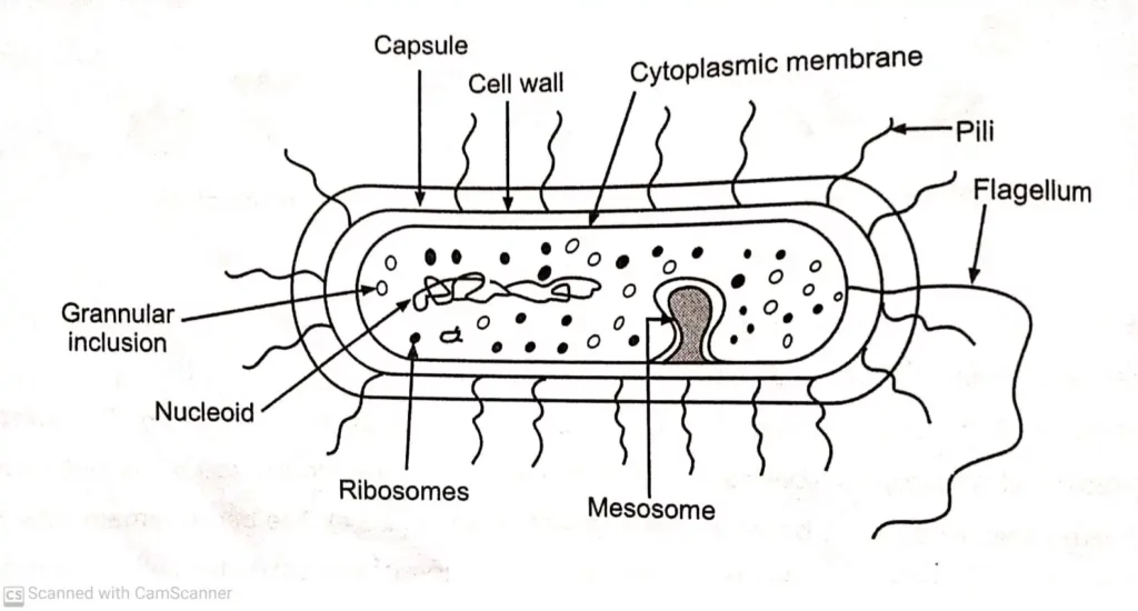
The structure of the bacterial cell is shown in PHOTO. The outer layer or cell envelope consists of two components a cell wall and a cytoplasmic or plasma membrane.Structure Parts and Functions of Bacteria Cell
Inside the plasma membrane, there is protoplasm comprising the cytoplasm, and cytoplasmic inclusions such as ribosomes, mesosomes, granules, vacuoles, and the nuclear body. The cell may be enclosed in a viscid layer, which may be a loose slime layer, or organized as a capsule.
Structure Parts and Functions of Bacteria Cell Just Full Read ?
Many bacteria have filamentous appendages called fimbriae or pili (organs of adhesion). Many bacteria also possess flagella which are organs of locomotion.
Flagella:
Flagella are long, slender, thin hair-like cytoplasmic appendages, which are responsible for the motility of bacteria. They are 0.01 to 0.02 um in diameter, and 3 to 20 um in length and are found in both Gram-positive and Gram-negative bacteria. These are organs of locomotion.
A large number of bacteria including a few coccal forms, most of the bacilli, and almost all of the spirilla and vibrios are motile using flagella.Structure Parts and Functions of Bacteria Cell
Flagella can be seen by an ordinary light microscope through special staining techniques in which their thickness is increased by mordanting. They can be easily visualized under an electron microscope and in some instances, they can be seen with dark ground microscopy.
The number and arrangement of flagella are characteristic of each bacteria. Flagella may be seen on the bacterial bodies in the following manner.
- Monotrichous: These bacteria have single polar flagellum eg. Vibrio cholera, Pseudomonas aeruginosa, and Spirillum.
- Lophotrichous: Bacteria have two or more flagella (tuft of flagella) only at one end of the cell e.g. Pseudomonas fluorescens.
- Amphitrichous: Bacteria have single polar flagella or tuft of flagella at both poles. e.g. Alcaligenes fecales, Aquaspirillum serpens
- Peritrichous: Several flagella present all over the surface of bacteria e.g. Escherichia coli and Salmonella typhi.
- Atrichous: Flagella is absent in these bacteria and hence they are non-motile e.g. Pasturella.
Flagella are made up of a protein (flagellin) similar to keratin or myosin and are responsible for bacterial motility. The motility of bacteria may be observed microscopically using the Hanging Drop technique or by detecting the spreading growth in a semi-solid agar medium.
Under the microscope, active motility has to be differentiated from the passive movements of the cells which is either due to air currents or Brownian movement.
Some helical bacteria (spirochetes) exhibit swimming motility in highly viscous media. However, they have flagella-like structures located within the cell, just beneath the outer cell envelope. These are called periplasmic flagella or endoflagella or axial fibrils e.g. Treponema pallidum.
Some bacterial species are motile only when they are in contact with a solid surface (e.g. Cytophaga species).
Bacterial Cell (Structure,Parts,Functions)
This type of motility is called gliding motility. The movement of a bacteria towards (positive) or away (negative) from a particular stimulus is called a taxi. The bacteria movement in response to a chemical stimulus is chemo-taxis and to light is photo-taxis.Structure Parts and Functions of Bacteria Cell
If a suspension of bacteria is placed on a slide under a coverslip, the aerobic bacteria accumulate at the edges of the coverslip while the anaerobic bacteria are at the center. It is called aero-taxis.
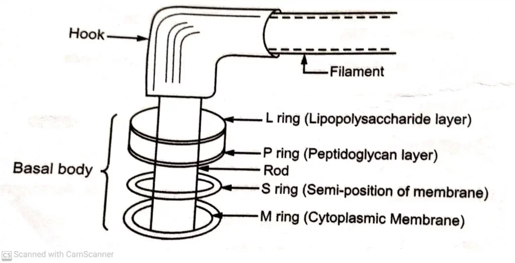
A flagellum has three basic parts i.e. (PHOTO) (1) Filament, (2) Hook, and (3) Basal body. The filament is the thin, cylindrical, extended outermost region with a constant diameter. The protein in the filament is made up of monomers called ‘flagellin’ with molecular weight ranging between 20,000 to 40,000.
The filament is attached to a slightly wider hook, consisting of a different protein. The basal body is composed of a small central rod inserted into a series of rings. Gram-negative bacteria contain four rings L-ring, P-ring, S-ring, and M-ring.
The L-ring is embedded in the Lipo-polysaccharide layer of the outer membrane, the P-ring in the Peptidoglycan layer, the S-ring is just above the cytoplasmic membrane (Semi-position of membrane), and the M-ring within the cytoplasmic Membrane. Gram-positive bacteria have only 5 and M rings in the basal body.
Pili or fimbriae:
Pili are hair-like microfibrils, 0.5 to 2 μm in length and 5 to 7 nm in diameter. They are thinner, shorter and more numerous than flagella (PHOTO).
They are present only on Gram-negative cells. Fimbriae are composed of a protein known as pilling and its molecular weight is 000 daltons. Fimbriae can be seen only under the electron microscope. They are related to motility and are found on motile as well as non-motile cells.
Structure Of Bacterial Cell with different Parts and Functions
They are best developed in freshly isolated strains and in liquid cultures. They may disappear after culturing on solid media.
Fimbriae and pili, these two terms are used interchangeably but they can be tinguished. Fimbriae can be evenly distributed over the entire surface of the cell or they cur at the poles of the bacterial cell. Each bacterium possesses 100 to 200 fimbriae. Pili are allies longer than fimbriae and number only one or two per cell.
They join bacterial cells in separation for the transfer of DNA (bacterial conjugation) from one cell to another cell and me, these pili are called sex pili or fertility pili (F-pili).
Structure Parts and Functions of Bacteria Cell
Functions of pili:
- Pili play a major role in attachment to surfaces. Hence pili is also called an organ of
- 2 Sex pili is used for the transfer of genetic material from the donor to the recipient
- Many fimbriated bacteria form surface pellicles in liquid media.
- Many fimbriated cells agglutinate red blood cells of guinea pigs, horses, fowl, pigs, sheeps and human beings e.g. Escherichia, Klebsiella. Hemagglutination provides a simple method for detecting the presence of such fimbriae.
- Fimbriae are antigenic. It is necessary to ensure that bacterial antigens employed for serological tests and preparation of antisera are devoid of fimbriae.
CAPSULE:
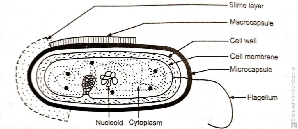
Many prokaryotic microorganisms synthesize amorphous organic exopolymers which are deposited outside the cell wall called capsules or slime layer or glycocalyx or sugar coat
The term capsule refers to the layer tightly attached to the cell wall while the slime layer is the loose structure that often diffuses into the growth medium (PHOTO), Capsule layer may be thin of a size less than 0.2 µm called microcapsule and thick layer of size more than 0.2 μm to 10 μm called macrocapsule.
Capsulated bacteria produce smooth colonies on surface of agar media. These bacteria are usually non-motile as flagella remains unfunctional in the presence of capsule.
Development of capsule is dependent on the existence of favourable environmental conditions such as sugar concentration, blood serum or growth in a living host.
Capsules may be composed of a complex polysaccharide (Klebsiella pneumoniae) or polypeptide (Bacillus anthracis) or hyaluronic acid (Streptococcus pyogenes) Water (98%) the main component of bacterial capsule. Capsule or slime layer has less affinity for basic dyes and is not visible in Gram staining. Special capsule staining techniques are used by using copper salts as mordants.Structure Parts and Functions of Bacteria Cell
Capsules may be easily observed by negative staining in wet films with Indian ink.
They are seen as clear halos around the bacteria against a black background. Capsular material is antigenic and may be demonstrated by serological methods. It is visualised by the reaction with a specific antibody which causes a characteristic swelling of the capsule. It is known as quelluing reaction.
Thin phenomenon is used for rapid identification of capsular serotypes. eg. Neisseria meningitidis, Streptococcus pneumonia , Hemophilus influenzae, Klebsiella, Yersinia and Bacillus species.
Functions of capsule:
A capsule serves a number of functions, depending on the bacterial species:
- Capsules protect the bacteria from antibacterial agents such as lytic enzymes.
- They inhibit phagocytosis (antiphagocytic) and contribute to the virulence pathogenic bacteria.
- They may provide protection against temporary drying by binding water molecules
- They may block attachment of bacteriophages.
- They may promote the stability of bacterial suspension by preventing the cells From aggregation and settling.
- They may promote attachment of bacteria to surfaces.(Structure Parts and Functions of Bacteria Cell)
Cell wall:
Cell wall is a rigid structure which gives definite shape to the cell, situated between the apsule and cytoplasmic membrane. It is about 10 – 20 nm in thickness and constitutes 0-30% of the dry weight of the cell. The cell wall cannot be seen by direct light microscopy nd does not stain easily by different staining reagents. It can be demonstrated by ifferential staining, plasmolysis, microdissection, electron microscopy and by using a pecific antibody.
The cell wall of bacteria contains diaminopimelic acid (DAP), muramic acid and teichoic acid. These substances are joined together to give rise to a complex polymeric structure Known as peptidoglycan or murein or mucopeptide. Peptidoglycan consists of three parts:
- A backbone composed of alternating N-acetyl glucosamine and N-acetyl muramic acid.
- A set of tetrapeptide side chains attached to N-acetylmuramic acid.
- A set of pentapeptide cross-bridges.
The backbone is same for all bacterial species. Tetrapeptide side chains and pentapeptide cross-bridges vary from species to species but the most commonly found side chain contains four amino acids as L-alanine, D-alanine, D-glutamic acid and diaminopimelic acid (DPA).Structure Parts and Functions of Bacteria Cell
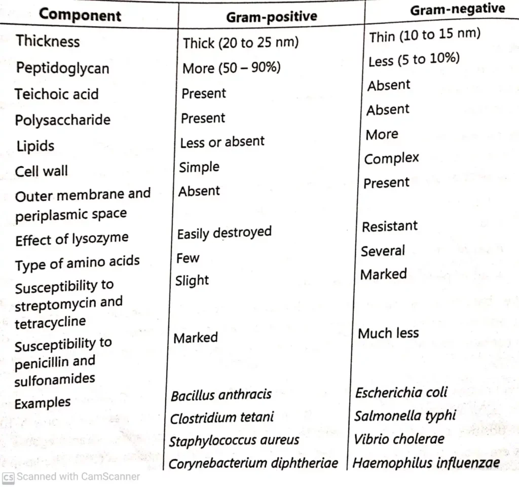
Peptidoglycan is the major constituent of the cell wall of Gram-positive bacterial (50 to 90%) whereas in the Gram-negative bacterial cell wall its presence is only 5-10%. Gram-positive cell wall also contains teichoic acid and polysaccharides. Gram-negative cell walls are more complex and the cell wall contains lipopolysaccharides (LPS). The lipopoly saccharides consists of three regions.
Region I is the polysaccharide portion determining the o-antigen specificity. Region II is the core polysaccharide. Region III is the glycolipid portion (Lipid A) and is responsible for endotoxic activities. The outermost layer of Gram-negative bacterial cell wall is called the outer membrane, which contains various proteins known as outer membrane proteins (OMP).
Among these are porins, which form transmembrane pores that serve as diffusion channels for small molecules. Differences between Gram positive cell wall and Gram-negative cell wall are given in Table.
Cell wall synthesis may be inhibited or interfered by many factors. Lysozyme enzyme present in many tissue fluids causes lysis of bacteria. If the Gram-positive cell is treated with lysozyme and the cell wall is completely removed, the cell is called a protoplast. If the Gram negative cell is treated with EDTA and the cell wall is partially removed (outer membrane), such a cell is called spheroplast.
The enzyme lysozyme is also capable of hydrolysing the B. 1-4 linkages between N-acetylmuramic acid and N-acetylglucosamine. Peptidoglycan is the site of action of a number of ß-lactam antibiotics such as penicillin, bacitracin, vancomycin and cycloserine Antibiotics which have a ß-lactam structure selectively inhibit the cell wall synthesis in bacteria.
First, the ß-lactam antibiotics bind to the cell receptors. These cell receptors are called as penicillin binding proteins (PBPs) which exist on the bacterial cell wall.
Some of these penicillin binding proteins are known to be transpeptidation enzymes. Different types of receptors are known to have different affinities to the ß-lactam drugs and have different effects to bring about the lysis of the cell. Gram-positive bacteria also have multiple transpeptidases but less than Gram-negative bacteria.
Endogenous autolytic enzymes (autolysins) are present in the cell wall and these are activated by ß-lactam antibiotics which cause lysis of cells. Autolytic enzymes are also able to hydrolyse peptidoglycan which is present in most bacterial cell walls.
Functions of cell wall:
- Cell wall is involved in growth and cell division of bacteria.
- It gives shape to the cell.
- It gives protection to the internal structure and acts as a supporting layer.
- It provides attachment to complement.
- It contains receptor sites for phages and colicin.
- It shows resistance to the harmful effects of environment.
Cytoplasmic membrane:
The cytoplasmic (plasma) membrane is a thin (5 to 10 nm) layer lining the inner surface of the cell wall and separating it from the cytoplasm. It is composed of phospholipids (20 to 30%) and proteins (60 to 70%). The phospholipids form a bilayer in which most of the proteins are tenaciously held and are called integral proteins (PHOTO).
These proteins can be removed only by destruction of the membrane with treatment by detergents. Other proteins are loosely attached and can be removed by mild treatments such as osmotic shock.
These proteins are called peripheral proteins. The phospholipid molecules are arranged in two parallel rows, called a phospholipid bilayer. Each phospholipid molecule contain a polar head composed of a phosphate group and glycerol. The non-polar tails are in the interior of the bilayer and the polar heads are on the two surfaces of the phospholipid bilayer.

Prokaryotic plasma membranes are less rigid than eukaryotic membranes due to lack of sterols. Electron microscopy shows the presence of three layers constituting a ‘unit membrane’ structure.
Functions of cytoplasmic (plasma) membrane:
- It acts as a semipermeable membrane controlling the inflow and outflow of metabolites to and from the protoplasm.
- It provides mechanical strength to the bacterial cell.
- It helps in DNA replication.
- It contains the enzyme, permease, which plays an important role in the passage of selective nutrients through membranes.
- It contains the enzymes involved in the biosynthesis of membrane lipids and various other macromolecules of the bacterial cell wall.
Cytoplasm:
The bacterial cytoplasm is a suspension of organic, inorganic solutes in a viscous wate solution. The cytoplasm of bacteria differs from that of higher eukaryotic microorganisms in not containing endoplasmic reticulum, Golgi apparatus, mitochondria and lysosomes.
I contains the nucleus, ribosomes, proteins and other water soluble components and reserve material. In most bacteria, extrachromosomal DNA (plasmid DNA) is also present.
Ribosomes:
The most notable structures in the bacterial cytoplasm are the ribosomes. They are involved in protein synthesis. Their number varies with the rate of protein synthesis (15,000/cell). The greater the rate of protein synthesis the greater the number of ribosomes.
These are ribonucleoprotein particles, which have a diameter of 200 A° and are characterised by their sedimentation properties. The bacterial ribosomes are referred to as 70S ribosomes (S-Svedberg unit, the unit of sedimentation). These ribosomes when placed in a low concentration of magnesium, dissociate into two components as 50S and 305 particles.
Each 50S particle contains one molecule of 23S-RNA, one molecule of 5S-RNA and 32 different proteins. The 30S subunit contains one molecule of 16S r-RNA and 21 different proteins. These ribosomes, during active protein synthesis are associated with the m-RNA and such associations are called polysomes.
Structure Parts and Functions of Bacteria Cell
Mesosomes:
In some bacteria, particularly in Gram-positive bacteria, depending upon the growth conditions the membrane appears to be infolded at more than one point. Such infoldings are called mesosomes. There are two types of mesosomes, i.e. central (septal) mesosome and peripheral (lateral) mesosome. Central mesosomes are present deep into the cytoplasm and located near the middle of the cell.
This mesosome is attached to bacterial chromosome and is involved in DNA segregation and in the formation of cross walls during cell division. Peripheral mesosomes are not restricted to a central location and are not associated with nuclear material. Mesosomes are also called chondroids and are visualised only under an electron microscope.
The presence of mesosome in large numbers have also been found in microorganisms that have a higher respiratory activity e.g. Azotobacter. In photosynthetic bacteria, the extent of membrane infolding has been related to pigment content and photosynthetic activity. In sporulating bacteria, the appearance of such infolding (mesosome formation) is a prerequisite for sporulation.
Intracytoplasmic inclusions:
Many species of bacteria produce cytoplasmic inclusion bodies which appear as round granules. They are characteristic of different species and depend on the condition of the culture. Volutin or metachromatic or Babes-Ernst granules are highly refractive, basophilic odies consisting of polymetaphosphate. They appear reddish when stained with olychrome methylene blue or toluidine blue.
Albert’s or Neisser’s special staining echniques are used for the study of volutin granules. Lipid granules consist mainly of -olymerised B-hydroxybutyric acid. They can be seen in unstained preparations and by taining with sudan black and by modified Ziehl-Neelsen stain. Polysaccharide granules can e stained with iodine. They are made-up of either glycogen (red brown) or starch (blue).
Nucleus:
Bacterial nucleus can be demonstrated by acid or ribonuclease hydrolysis. They may be seen by a light microscope after staining (Feulgen stain) or by electron microscopy.
They appear as oval or elongated bodies, generally one per cell. The genome consists of a single molecule of double stranded DNA arranged in a circle. It may open under certain conditions to form a long chain about 1000 μm in length. Bacterial nucleus does not possess nuclear membrane, nucleolus and deoxyribonucleoprotein.
The bacterial chromosome is haploid and replicates by simple fission instead of mitosis as in a eukaryotic cell.
A bacterial cell may posses extranuclear genetic elements consisting of DNA. These cytoplasmic carriers of genetic information are termed as plasmids or episomes. They may be transmitted to daughter cells during binary fission or by conjugation. Presence of plasmid confers the characteristics of drug resistance, conjugation ability, pathogenesis and nitrogen fixation ability.
Spores:
Many bacterial species produce spores inside the cell (endospores) as well as outside the cell (exospores). e.g. Bacillus anthracis, Bacillus subtilis, Clostridium tetani, Clostridium botulinum, Coxiella burnetii, Sporosarcina, Thermoactinomycetes, Streptomyces, Actinopolyspora species etc.
Endospores are thick-walled, highly refractile bodies that are produced one per cell. Each bacterial spore, on germination forms a single vegetative cell.
Therefore, sporulation in bacteria is a method of preservation and not reproduction. Spores are extremely resistant to dessication, staining, disinfecting chemicals, radiation and heat. They remain viable for centuries and help bacteria to survive for long periods under unfavourable environments. Spores of all medically important bacteria are destroyed by moist heat sterilisation (autoclave) at 121°C for 20 minutes.
All endospores contain large amount of dipicolinic acid (DPA) with 10 to 15 percent of the spores being dry weight. It occurs in combination with large amounts of calcium, which is present in the central part of the spore (core). The calcium – DPA complex may possibly play a role in the heat resistance of endospores. Spore can be detected by staining with a specially prepared stain (Schaeffer-Fulton method) along with heat.
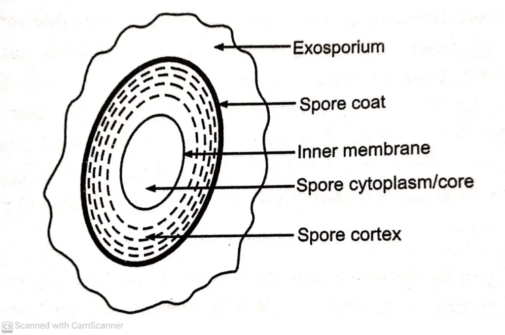
The endospore consists of a core or envelope or protoplast. The protoplast contains the DNA and components of protein synthesis like ribosome, t-RNA and enzymes. The spore envelope consists of the inner membrane, cortex, outer membrane and spore coat. In some species the outer layer also develops exosporium which bears ridges and folds (PHOTO). The multilaminated cortex develops between the inner and the outer membrane. It consists of peptidoglycan. The spore coat is formed outside the outer membrane and is made up of keratin-like protein. They are unaffected by chemical treatment and confer the resistance to spore from chemicals.
Structure Parts and Functions of Bacteria Cell
The spores may be round, oval or elongated occupying a terminal, subterminal or central position. They may be narrower than the width of the bacilli and bulging.

